


The following research papers highlight the impact that Irlen Syndrome / SSS has on brain function and anatomy.
The most current research on Irlen Syndrome / SSS and the use of colour utilises advanced brain-mapping technology to show actual changes and normalisation of brain functioning that is not achieved through ophthalmological treatments (plain lenses, prisms, or vision therapy). Researchers have utilized functional magnetic resonance imaging (fMRI), visual evoked responses (VER), and single photon emission computed tomography (SPECT) scans to objectively document the profound effects of visual sensory overload on the brain and the normalisation of brain activity when individually-prescribed, precision-tinted coloured filters are worn.
A Magnetoencephalographic Investigation of Visual Information Processing in Irlen’s Scotopic Sensitivity Syndrome
by Jeffrey Lewine, Ph.D., John Davis, Ph.D., Sherri Provencal, M.A., James Edgar, M.A., and William Orrison, Jr., M.D.
Lewine et al. (1997) utilized magnetoencephalography (MEG) to characterise visual responses in conditions with and without lenses. In all cases, the evoked magnetic signal reflected a complicated pattern of bilateral activation of multiple cortical generators. A major difference in with and without lens conditions was seen between 170 and 200 msec post-stimulus. The data suggests that the coloured Irlen lenses provide for normalisation and crystallisation of visual information processing in individuals with Irlen Syndrome.
Visual Revoked Responses: Yellen-Schweller Effect
by Drew Yellen
Yellen and Schweller (2009) utilised state-of-the-art Visual Evoked Responses (VER), a portion of their comprehensive neuroelectrical evaluation of patients called the DESA®, and discovered that individuals with Irlen Syndrome have early hyper reactivity to visual stimuli somewhere between 30-60 milliseconds, and it is 3-9 standard deviations above normal (the Yellen-Schweller Effect). Irlen Spectral Filters reduce the standard deviation abnormalities of the Yellen-Schweller Effect, lessening of the delay of the brain coming back “online” and allowing it to clear sooner.
Toward an Authentic Diagnostic Impression Using Clinical Composites and Functional Brain Imaging for an Improved Understanding of Irlen Syndrome
by Robert Dobrin, M.D., F.A.A.P
Light and the Brain
Brain in the News Newsletter by Daniel G. Amen M.D.
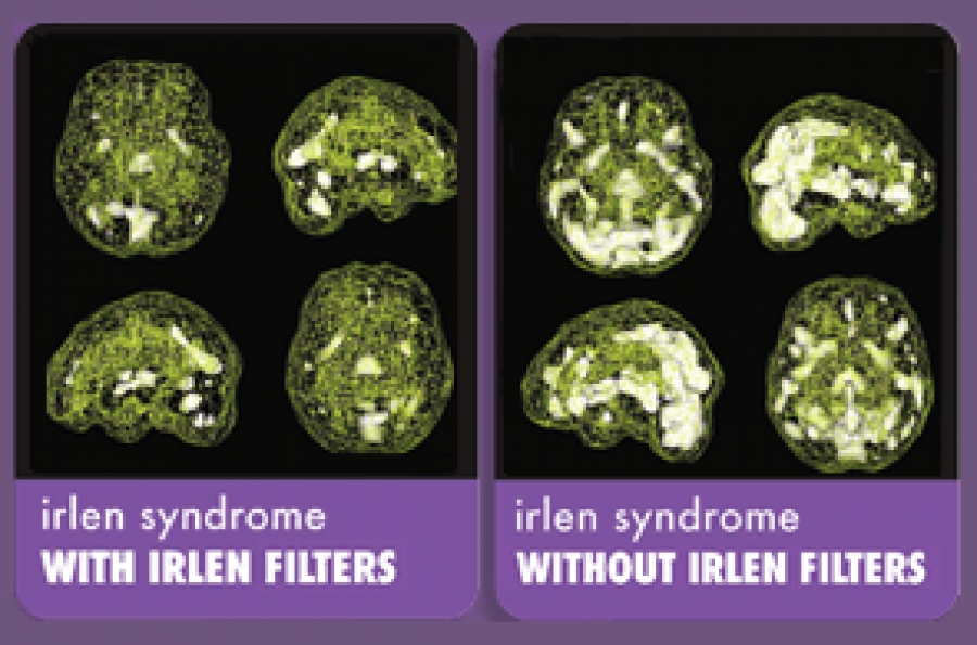
In one study by Amen and colleagues, comparing the brains of 42 people with Irlen syndrome to 200 age-matched individuals without any evidence of Irlen syndrome, SPECT scans showed increased activity in the brain’s emotional and visual processing centres and decreased activity in the cerebellum (an area that helps to integrate coordination and new information).
A Functional Neuroimaging Case Study of Meares-Irlen Syndrome/Visual Stress (MISViS)
Chouinard, B.D., Zhou, C.I., Hrybouski, S., Kim, E.S., Cummine, J. (2012).) Brain Topography, 25 (3), pp. 293-307. http://link.springer.com/article/10.1007%2Fs10548-011-0212-z#page-1
Chouinard et al. (2011) compared the neurological characteristics of a person with Irlen Syndrome with control subjects who were participating in a language. The descriptive results indicated that there are numerous significant differences in many areas of the brain cortex between the control subjects and the individual with Irlen Syndrome, providing evidence of a neurobiological foundation to Irlen Syndrome.
fMRI Evidence that Precision Ophthalmic Tints Reduce Cortical Hyperactivation in Migraine
Huang, J., Zong, X., Wilkins, A., Jenkins, B., Bozoki, A., Cao, Y. (2011).. Cephalagia, 31(8):925-36. http://www.ncbi.nlm.nih.gov/pmc/articles/PMC3132147/
Huang et al. (2011) used fMRI to investigate differences between individuals suffering visual stress and controls in relation to migraine and to determine the effectiveness of precision-tinted coloured filters for individuals suffering from visual stress. The research showed a normalisation of cortical activation and spatial frequency tuning in the migraineurs by precision tinted filters that suggests a neurological basis for the therapeutic effect of these lenses in reducing visual cortical hyperactivation in migraine.